Scientists have identified a new mechanism that accelerates aging in the brain and gives rise to the most devastating biological features of Alzheimer’s disease.
The findings also unify three long-standing theories behind the disease’s origins into one cohesive narrative that explains how healthy cells become sick and gives scientists new avenues for screening compounds designed to slow or stop disease progression, something existing medications cannot do.
“We now have a better understanding of the molecular factors that lead to Alzheimer’s disease, which we can leverage to develop improved and desperately needed treatment and prevention strategies,” said Viviane Labrie, Ph.D., an assistant professor at Van Andel Research Institute (VARI) and senior author of the study, which appears in the May 21 edition of Nature Communications. “Alzheimer’s is a major growing public health problem around the world. We need better options for patients and we need them soon.”
Alzheimer’s disease is the sixth leading cause of death in the U.S. and the most common cause of dementia worldwide. An estimated 5.8 million people in the U.S. and 44 million people worldwide have Alzheimer’s. By 2050, those numbers are expected to rise to 14 million and 135 million respectively, due in part to a growing and aging global population.


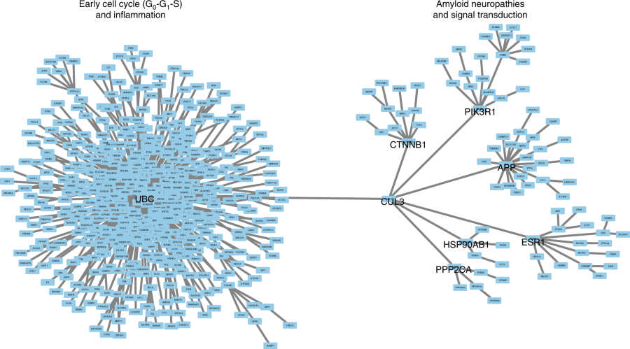
The findings center on genetic “volume dials” called enhancers, which turn the activity of genes up or down based on influences like aging and environmental factors. Labrie and her colleagues took a comprehensive look at enhancers in brain cells of people at varying stages of Alzheimer’s and compared them to the cells of healthy people. They found that in normal aging, there is a progressive loss of important epigenetic marks on enhancers. This loss is accelerated in the brains of people with Alzheimer’s, essentially making their brain cells act older than they are and leaving them vulnerable to the disease.
At the same time, these enhancers over-activate a suite of genes involved in Alzheimer’s pathology in brain cells, spurring the formation of plaques and tangles, and reactivating the cell cycle in fully formed cells — a highly toxic combination.
“In adults, brain cells typically are done dividing. When enhancers reactivate cell division, it’s incredibly damaging,” Labrie said. “The enhancer changes we found also encourage the development of plaques, which act as gasoline for the spread of toxic tangles, propagating them through the brain like wildfire. Taken together, enhancer abnormalities that promote plaques, tangles and cell cycle reactivation appear to be paving the way for brain cell death in Alzheimer’s disease.”
Importantly, Labrie and her colleagues linked enhancer changes to the rate of cognitive decline in Alzheimer’s patients.
The study is the first comprehensive investigation of enhancers in human brain cells and in Alzheimer’s disease, and included in-depth analysis of epigenetic, genetic, gene expression and protein data.
Next, the team plans to develop new experimental systems to screen compounds that may fix dysregulation in enhancers and that have potential as new treatments or preventative measures.

